This
information website is focused
on assisting anyone looking to
better understand how their body
functions, at the cellular
level, and how mitochondria play
a
critical role in both health
and longevity as well as disease
and aging. Some will need an
introductory overview while
others may be further advanced
in their knowledge of this
critical organelle called the
Mitochondria.
For a laymens perspective, you
will find sections that will
provide videos and graphics that
are provided with the intent of
generating VISUAL AIDS that will
be imprinted in your mind long
after viewing this website.
These VISUAL AIDS & the data
provided will show you the many
roles that Mitochondria play as
the KEY POWER GENERATOR of each
cell.
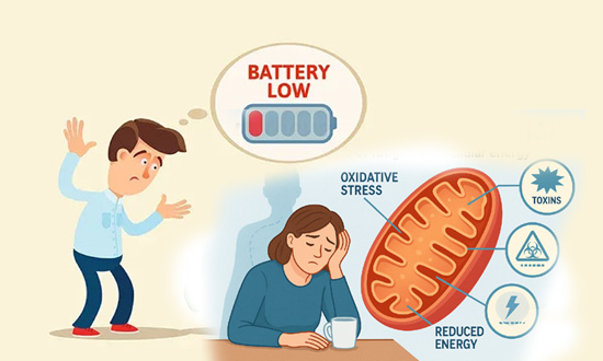
IT IS ALL ABOUT THE BATTERY
Cars, Phones and
Flashlights are all
dependent on Batteries
to work.
A dead battery results
in a
CAR that won't start,
a PHONE that won't turn
on and
a FLASHLIGHT that will
not give off any light.
Mitochondria
function as the Battery
of every Cell
If your mitochondria are
dysfunctional,
each cell and
each organ in
your body is operating
at a limited capacity.
Cars
struggle to start
without a "Good
battery"
Cells
struggle to
function without
"Good
Mitochondria"
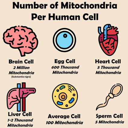
What Organs &
Tissue have the most
cellular Mitochondria
.
The
number of mitochondria
per organ varies
drastically based on energy
needs,
with high-demand
organs like the heart,
liver, muscles, and
brain having thousands
per cell (heart muscle
cells can have
5,000-8,000),
while other cells have
fewer, and mature red
blood cells have none,
as the quantity reflects
the cell's energetic
workload, with
mitochondria generating
ATP energy.
High-Energy Organs
(Thousands per cell)
Heart
Heart muscle cells are packed with
mitochondria (5,000-8,000 per cell)
to power constant contraction.
Liver
Liver cells have many mitochondria
(over 2,000) for metabolic functions,
drug detoxification, and waste
breakdown.
Muscles
Muscle cells, especially skeletal
and cardiac,
have
abundant mitochondria
for movement and sustained activity.
Brain
Neurons require immense energy, with
some estimates
suggesting up to 2 million
mitochondria per neuron,
as the brain is highly
energy-intensive.
Lower-Energy Cells
.
Cells in less active tissues, or
those with specialized functions
like oxygen transport (red blood
cells),
have significantly fewer
mitochondria, or none at all.
Why the Difference?
Energy Demand:
Mitochondria are the cell's
powerhouses, producing ATP (energy).
Cells that perform more work, like
pumping blood (heart) or processing
nutrients (liver),
need more mitochondria.
Volume:
In some cases, like liver cells,
mitochondria can make
up a significant portion (around
20%) of the cell's volume.
Advanced Video
Beginners
Video
Mitochondria explained in 6 minutes
What
are mitochondria and how do they
fuel the body?
"Mitochondrial
Function" determines your State of Health?
Efficient
Mitochondrial metabolism (of the Batteries
in your cells) is critical for
providing the
proper levels of energy so that genes and proteins
can be properly transcribed. Without
efficent Mitochondria, the
metabolic rate of a cell is severely diminished.
Low Batteries = Low Power. Without the
proper levels of energy (Electricity), errors in gene and
protein transcription can occur. .
Disease State vsHealthy State
Without Mitochondrial Support? Very Low
energy levels = Possible Genetic
Errors
DISEASE STATE WITHIN THE
CELL
With
Metabolically Targeted Therapy
Optimizes energy levels = Proper Gene &
Protein Transcription
HEALTHY STATE WITHIN THE
CELL
Metabolic
dysfunction as a result of Mitochondrial
Dysfunction is at the heart of a multitude of
clinical conditions, including cancer. The
approach of implementing products or
compounds that increase or maintain
mitochondrial function thereby combatting metabolic dysfunction can be
viewed as a metabolically targeted therapy
(MTT).
Has Clinical Research been
looking
in the wrong place
for the past 50 years? The 3rd party
clinical studies found on this site suggest an emphatic,
. “YES!”
After reading the 3rd party peer reviewed
articles
listed below and posted throughout this website, one central theme
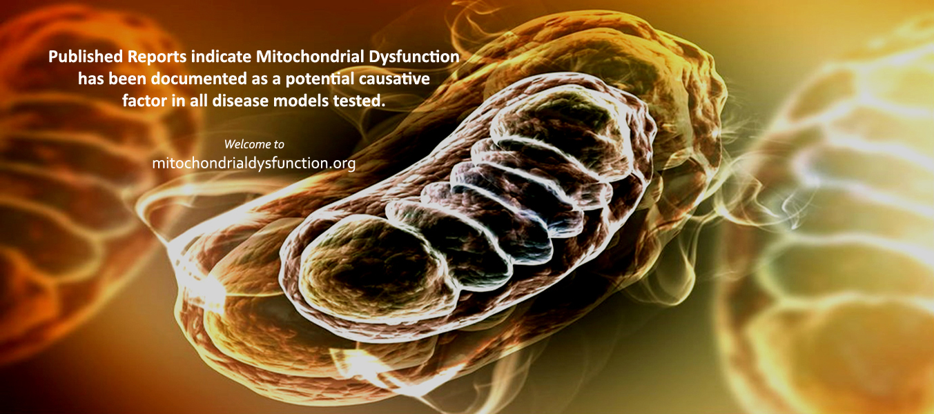
becomes clear. . "Mitochondrial dysfunction has been implicated in
nearly all pathologic and toxicologic conditions."
During hypoxia, the HIF-1 gene
turns on hypoxic dependent genes
and represses or turns off normoxic
dependent genes
The latest research suggests that the
Human Cell contains as many as 20,000
different genes but only a fraction of
these genes are turned on at one time.
THE HEALTHY CELL:
When proper oxygen levels are
available under normoxic
conditions, hypoxic genes, like HIF-1,
are repressed, or turned off
resulting in the homeostasis of a
healthy cell and normal mitochondrial
function.
THE DISEASED CELL:
However, when a cell becomes
hypoxic, HIF-1 levels and other
hypoxic dependent genes are transcribed
resulting in a disease state within the
cell.Increased HIF-1
levels results in the downstream
transcription of Vascular Endothelial
Growth Factor
(VEGF) – a promoter of angiogenesis;
Glucose Transport 1
(GLUT1) and glycolytic enzymes
– critical components in anaerobic
respiration;
and Erythropoietin
(EPO) – responsible for the
differentiation of red blood cells.
(supporting article: Free Radic Biol Med. 2009 Jan)
The transcription
of these hypoxic dependent genes would
have never occurred if the cell had
remained under normoxic conditions.
This cellular response to low oxygen, or
hypoxia, involves the regulation of many
cellular pathways that shut down low
priority cellular activity and increase
stress responses.
These signals from the
environment
during hypoxia
activate various proteins which
are called transcription factors.
These proteins bind to
regulatory regions of a gene and
increase or decrease the level of
transcription. By controlling
the level of transcription, this process
can determine the amount of protein
product that is made by a gene at any
given time.
. THE HUMAN CELL &
OXIDATIVE STRESS Transition from
"Healthy State" to "Stressed State" to "Disease
State" .
The Human cell exists in
1 of 3
different states at all times
Either Cell maintains proper Oxygen levels orHIF-1α
is upregulated which alters gene transcription! .
HEALTHY STATE
Normal oxygen levels
HIF-1 levels are degraded & remain low Stress&Disease genesstayturned off
CELL STATUS
Cellular function/Gene Transcription is optimized
STRESSED STATE
Reduced oxygen levels
oxidative stress begins to accumulate Stress genes
are upregulated/Healthy genes
are down-regulated
CELL STATUS
Increased oxidative stress = endogenous antioxidant response
DISEASE STATE
Significantly reduced oxygen levels
long term oxidative stress/ HIF-1 levels
increase Disease genesactivated/Healthy genes
are turned off
CELL STATUS
The cell is suffocating - survival
is at risk
COMMON SENSES QUESTIONS
Why have
scientists spent the last 50 years
trying to repair genes
AFTER
the cell has entered
the disease state
when
the negative"transcribing
factor" is known to be
hypoxia/oxidative stress
which mitigates mitochondrial function?
Wouldn't it
make more sense to implement
strategies to prevent hypoxia &
reverse oxidative stress thereby
preventing
the
transcription of each of these
hypoxic dependent genes
that lead to a disease state within
the cell?
Why are these
same scientists suggesting that
disease is based on GENETIC
ERRORS when most of these
errors
occur due to
the transcription of genes that are
only
transcribed/turned on under hypoxic
conditions?
ANSWER
If
mitochondrial dysfunction is
associated with oxidative stress & the upregulation
of HIF-1, the only sensible response
would be to
develop compounds
that can reverse
this dysfunction
and
prevent
hypoxia from occurring.
.
When intracellular ROS
overwhelms the Cell The cell enters the "Stressed State"
leading potentially, to the "Disease State"
.
The delicate balance between
intracellular oxidation and antioxidation is
critical in order to maintain proper gene
transcription. Under physiological Oxidative Stress
conditions, the human antioxidative defense system,
which includes superoxide dismutase
(SOD), catalase (CAT), glutathione peroxidase (GPx),
glutathione (GSH) alpha lipoic acid(ALA) and
coenzyme Q10(CoQ10), must efficiently
reduce excess
reactive oxygen species (ROS) like
superoxide anions (O2.-), hydroxyl
radicals (OH), alkoxyl radicals (RO) and
peroxyradicals (ROO). .
This ongoing day to day, year to year, maintenance of cellular respiration via optimal
intracellular oxygen levels dictates the health of
every cell in the human body. Under
various conditions, the efficiency of our endogenous
antioxidative defense system may be diminished
resulting in increased levels of intracellular
oxidative stress. If this oxygen deficient state is
not reversed, adeficient antioxidant defense system can become
overwhelmed which can lead to a
disease state within the cell. This
disease state may persist due to
continuous altered gene transcription.
.
During each Oxidative Stress
event the ability of cells to adapt to low oxygen levels is
essential for processes such as development,
growth, metabolism, and
angiogenesis. However, the response to a decrease
in oxygen supply, referred to as hypoxia,
is also involved in numerous human diseases
including cancer, inflammatory
conditions, and vascular disease.
. Thehypoxia-inducible factor 1-α (HIF-1α)
is a key player in the cells response to
hypoxia
and is
kept under stringent
regulation.
.
During normoxia(normal oxygen
levels), the levels of
HIF-1α
stay low due to degradation by the
ubiquitin-proteasome system. However, in
response to hypoxia(low oxygen levels),
the degradation is blocked and HIF-1α levels
increase in order
to promote a
transcriptional response essential for
proper adaptation
and
survival during stress or disease states within each
cell.
.
Oxidative Stress can lead
to The upregulation of HIF-1
which initiates Mitochondrial Dysfunction
.
Most
causes of mitochondrial dysfunction tend to involve
oxidative stress which can be generated by a myriad
of sources. These levels of oxidative stress can be
dramatically increased and persist at dangerous
levels if the human body is continually exposed to
more than 1 of the following sources at the same
time......see list below.
Exposure to
these sources
can lead to Mitochondrial
Dysfunction
.
alcohol, artificial trans fats,
aspirin, excess calories, glucocorticoids,
homocysteine, iron overload, lipid peroxidation,
lipopolysaccharide, MSG, nutrient deficiencies,
oxidized LDL, pro-inflammatory cytokines,
prescription drugs, sleep deprivation, smoking, statins
and toxic heavy metals.
Per the published studies found on this
website, the human body is constantly exposed to
many of these known causative factors resulting in Oxidative
stress and the upregulation of HIF-1. As
noted in the article below from Johns Hopkins, if this
dysfunction is not reversed, it is
only a matter of time before this long term
oxidative stress will manifest itself in disease and
suffering.
Hopkins
researchers discover unsuspected
genetic switch (HIF-1) that turns off
Mitochondria
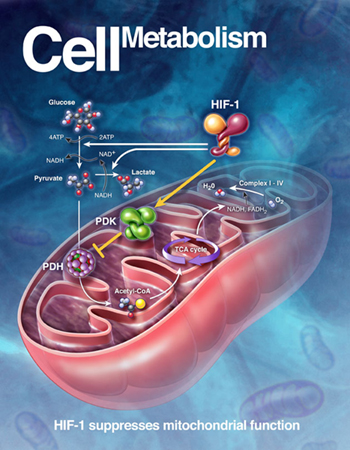
. HIF-1 suppresses
mitochondrial function
A cell’s energy demands are
met by two major types of
sugar (glucose) using
machines similar to the 2
types of engines in a hybrid
car. One machine,
the mitochondrion, is an
organelle that breaks down
the glucose-using oxygen and
produces ATP. The
other does the same thing -
albeit less efficiently -
without using oxygen,
in a process called
glycolysis.
Like the hybrid car, cells use
oxygen and the internal
combustion engine at higher
speeds and rely on an electric
engine without need for oxygen
consumption at lower speeds.
Cells
consume glucose through its main
energy-producing machine, the
mitochondrion, when oxygen is
ample. But like the
internal combustion engine, this
process generates pollutants or
toxic oxygen molecules.
At lower oxygen levels, when
cells are starved for oxygen -
as during exertion or trauma -- the genetic switch that
the Hopkins researchers found
deliberately shuts off the
cell’s mitochondrial combustion
engine, which
scientists had long - and
erroneously -- believed ran
down on its own due to lack of
oxygen.
“The unexpected
discovery is that this genetic
switch actively shuts off the
mitochondrion under low oxygen
conditions, apparently to
protect cells from mitochondrial
toxic oxygen pollutants,” said Chi Van Dang,
M.D., Ph.D., professor of
medicine, cell biology, oncology
and pathology, and vice dean for
research at the Johns Hopkins
University School of Medicine.
Dang says the switch may be a target for
cancer drugs because a cancer cell’s
survival depends on it to convert
glucose to lactic acid through
glycolysis even in the presence of ample
oxygen. Disruption of the
switch(HIF-1) by a drug may cause cancer
cells to pollute themselves with toxic
oxygen molecules and undergo apoptosis
or cell death.
The disruption of this link
blocks the tendency of
the mitochondrion to make toxic
molecules
as it struggles to produce ATP
during hypoxia.
These toxic molecules,
called reactive
oxygen species (ROS),
damage molecules
in the cell and even
cause the cell to
undergo apoptosis.
"But our discovery clearly shows that
hypoxia doesn’t simply trigger a passive
shutdown of the mitochondrion,” said
Dang. “Instead, HIF-1 acts as a
genetic switch to actively shut down
mitochondrial function and prevent the
production of reactive oxygen species.”
Relationship between oxidative stress
and HIF-1 alpha
mRNA during sustained hypoxia in humans.
Abstract
The aim of this study was to investigate
the relations among reactive
oxygen species (ROS),
hypoxia inducible factor (HIF-1
alpha) gene expression,
HIF-1 alpha target gene
erythropoietin (EPO), and
vascular endothelium growth
factor (VEGF) in humans.
Five healthy men (32+/-7 years,
mean+/-SD) were exposed to 12 h of
sustained poikilocapnic hypoxia
(P(ET)O(2)=60 mmHg).
DNA oxidation (8-hydroxy-2'-deoxyguanosine,
8-OHdG), advanced oxidation protein
products (AOPP), EPO, and VEGF were
measured in plasma and HIF-1 alpha mRNA
was assessed in leukocytes before and
after 1, 2, 4, 6, 8, 10, and 12 h of
exposure to hypoxia. HIF-1 alpha mRNA
amount increased during the first two
hours of hypoxic exposure and then
returned to baseline levels. The
findings reveal an up-regulation of
HIF-1 alpha (+68%), VEGF (+46%), and EPO
(+74%). AOPP increased continuously from
4 h (+69%) to 12 h (+216%) of hypoxic
exposure while 8-OHdG increased after 6
h (+78%) and remained elevated until 12
h.
During the "acute" increase
phase of HIF-1 alpha (between 0
and 2 h), 8-OHdG was positively
correlated with HIF-1 alpha
(r=0.55). These findings suggest that
hypoxia induces oxidative stress
via an overgeneration of reactive oxygen
species (ROS).
Finally, this study in humans
corroborates the previous in vitro
findings demonstrating that ROS
is involved in HIF-1 alpha
transcription.